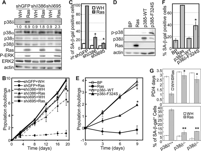
Fig 1.
p38δ mediates oncogenic ras-induced senescence. (A) Western blot analysis of BJ cells transduced with shRNA for GFP (shGFP) or p38δ (shδ386 and shδ695) and Ha-rasV12 (Ras) or vector (WH) on day 8 after ras transduction. Numbers represent relative levels of protein. (B) Population doublings of BJ cells transduced with shRNA for GFP (shGFP) or p38δ (shδ386 and shδ695) and Ha-rasV12 (Ras) or vector (WH) over 19 days starting at PD 30. Values are means ± SDs for triplicates. *, P < 0.05 versus shGFP by Student's t test. (C) Percentage of SA-β-Gal-positive cells in BJ populations transduced with shRNA for GFP (shGFP) or p38δ (shδ386 and shδ695) and Ha-rasV12 (Ras) or vector (WH). Values are means ± SDs for triplicates. *, P < 0.005 versus shGFP by Student's t test. (D) Western blot analysis of BJ cells transduced with Ha-rasV12 (Ras), wild-type p38δ, the p38δ-F324S mutant, or vector (BP) on day 8 posttransduction. (E) Population doublings of BJ cells transduced with Ha-rasV12 (Ras), wild-type p38δ, the p38δ-F324S mutant, or vector (BP) over 9 days starting at PD 30. Values are means ± SDs for triplicates. *, P < 0.05 versus BP by Student's t test. (F) Percentage of SA-β-Gal-positive cells in BJ populations transduced with Ha-rasV12 (Ras), wild-type p38δ, the p38δ-F324S mutant, or vector (BP). Values are means ± SDs for triplicates. *, P < 0.05 versus BP by Student's t test. (G) p38δ deficiency disrupts ras-induced senescence in MEFs. p38δ+/+ and p38δ−/− MEFs were transduced with Ha-rasV12 or vector (WH). A total of 2 × 10⁴ cells were seeded into 12-well plates on day 5 after ras transduction, after selection of transduced cells. Cells were counted 4 days after seeding, and PD was calculated (top graph). Cells were stained for SA-β-Gal on day 10 after ras transduction. The percentage of cells positive for SA-β-Gal was quantified (bottom graph). Values are means ± SDs for duplicates. *, P < 0.05 versus p38δ+/+; **, P < 0.005 versus p38δ+/+ (Student's t test).

