Fig 3.
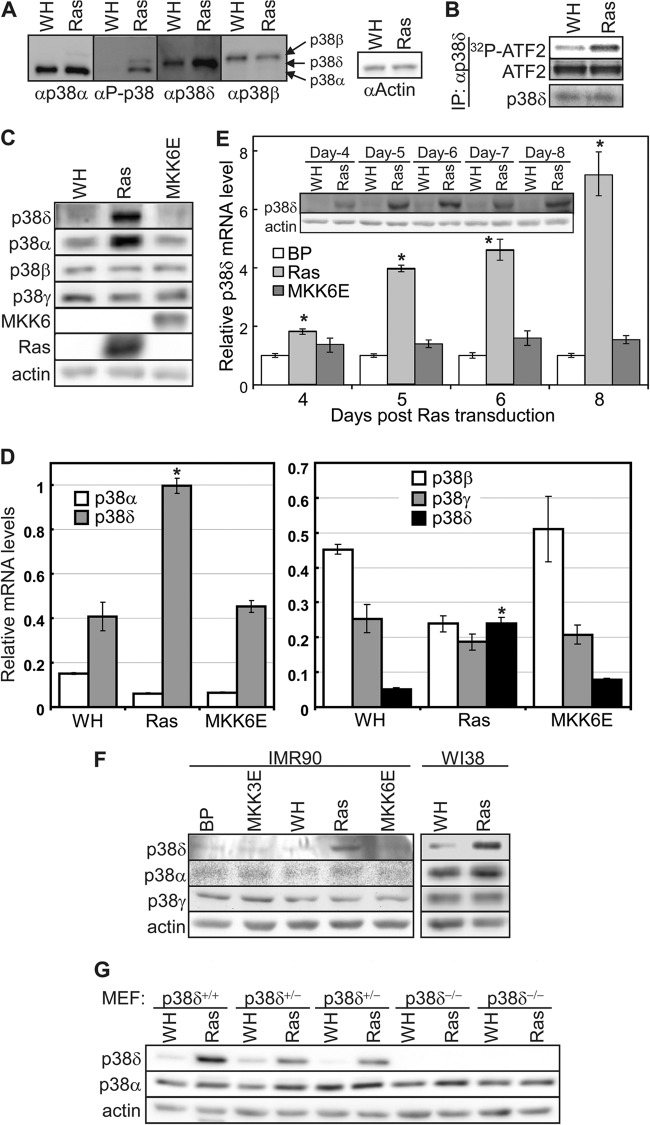
Oncogenic ras induces p38δ activity and expression. (A) Western blot analysis of BJ cells transduced with Ha-rasV12 (Ras) or vector (WH). Identical sets of lysates were resolved side by side on the same SDS-PAGE gel and transferred to a nitrocellulose membrane. The membrane was cut into pieces, each containing one set of lysates, which were then hybridized to the antibody against phospho-p38, p38α, p38δ, and p38β, respectively. The enhanced chemiluminscence signals were captured after the membranes were realigned into the original position. The positions of p38α, p38δ,and p38β are marked by arrows. (B) Oncogenic ras induces the kinase activity of p38δ. An equal amount of p38δ was immunoprecipitated from BJ cells transduced with Ha-rasV12 (Ras) or vector (WH) after adjustment of the input and assayed for kinase activity toward ATF2. Part of the IPs was subjected to Western blotting to ensure an equal amount of p38δ IP. (C) Western blot analysis of the protein levels of p38 isoforms in BJ cells transduced with Ha-rasV12, MKK6E, or vector (WH) on day 8 after ras transduction. (D) Analysis of the mRNA levels of p38 isoforms in BJ cells transduced with Ha-rasV12, MKK6E, or vector (WH) on day 8 after ras transduction, by quantitative real-time RT-PCR. Signals for p38 were normalized to that of PBGD. Values are means ± SDs for triplicates. *, P < 0.01 versus WH by Student's t test. (E) Time course analysis of the p38δ mRNA (bar graph) and protein (inset) levels in BJ cells transduced with Ha-rasV12, MKK6E, or vector (BP) on day 4 through day 8 after ras/MKK6E transduction. Bar graph, signals for p38δ mRNA were detected by quantitative real-time RT-PCR and normalized first to that of PBGD and then to that from cells transduced with vector control (BP). Values are means ± SDs for triplicates. (Inset) The p38δ and actin protein levels were detected by Western blotting. *, P < 0.05 versus BP by Student's t test. (F) Western blot analysis of p38 isoforms in IMR90 and WI38 cells transduced with Ha-rasV12, MKK3E, MKK6E, or vector (WH or BP) on day 8 after ras/MKK3/6E transduction. (G) Western blot analysis of p38 isoforms in p38δ+/+, p38δ+/−, and p38δ−/− MEFs transduced with Ha-rasV12 (Ras) or vector (WH) on day 8 after ras transduction.

