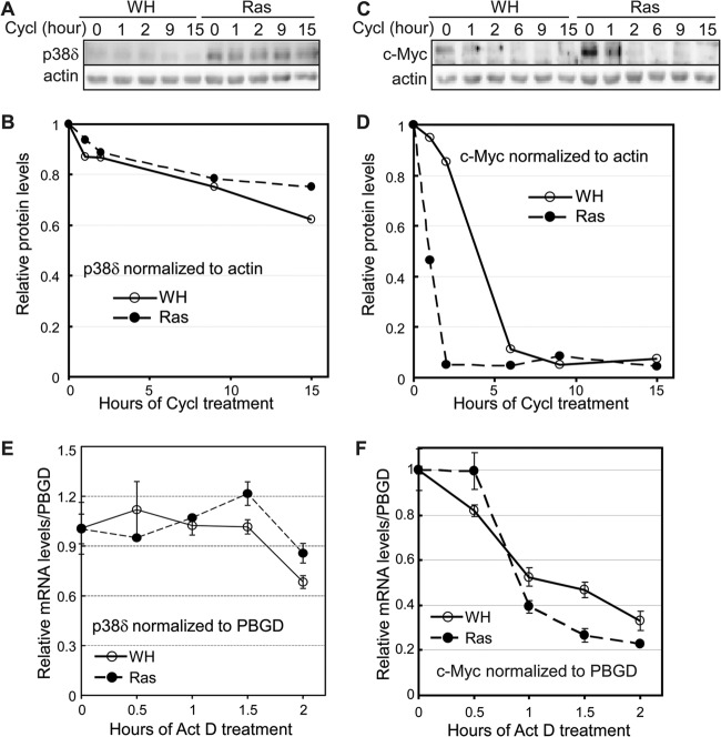
Fig 5.
Oncogenic ras does not stabilize p38δ protein or mRNA. (A) Western blot analysis of p38δ protein levels in BJ cells transduced with Ha-rasV12 (Ras) or vector (WH) after treatment with cycloheximide (Cycl) for 0, 1, 2, 9, or 15 h on day 8 after ras transduction. (B) Quantification of relative p38δ protein levels in panel A. The relative p38δ protein level was calculated by dividing the p38δ signal at each time point after cycloheximide treatment by that at 0 h, after normalization to the actin signal. (C) Western blot analysis of c-Myc protein levels in BJ cells transduced with Ha-rasV12 (Ras) or vector (WH) after treatment with cycloheximide for 0, 1, 2, 6, 9, or 15 h on day 8 after ras transduction. (D) Quantification of relative c-Myc protein levels in panel C. The relative c-Myc protein level was calculated by dividing the c-Myc signal at each time point after cycloheximide treatment by that at 0 h, after normalization to the actin signal. (E) Quantification of p38δ mRNA levels by real-time RT-PCR in BJ cells transduced with Ha-rasV12 or vector (WH), after treatment with actinomycin D for 0, 0.5, 1, 1.5, or 2 h on day 8 after ras transduction. The relative p38δ mRNA level was calculated by dividing the p38δ signal at each time point after actinomycin D treatment by that at 0 h, after normalization to the PBGD signal. Values are means ± SDs for triplicates. (F) Quantification of c-Myc mRNA levels by real-time RT-PCR in BJ cells transduced with Ha-rasV12 or vector (WH) after treatment with actinomycin D for 0, 0.5, 1, 1.5, or 2 h on day 8 after ras transduction. The relative c-Myc mRNA level was calculated by dividing the c-Myc signal at each time point after actinomycin D treatment by that at 0 h, after normalization to the PBGD signal. Values are means ± SDs for triplicates.

