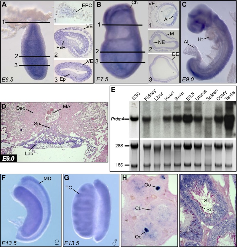
Fig 1.
Prdm4 RNA expression analysis during embryonic and postnatal development. (A) Prdm4 whole-mount in situ hybridization at E6.5 shows widespread expression in all tissues, with the exception of the EPC. (B) At E7.5, Prdm4 expression is apparent throughout the embryonic and extraembryonic tissues. (C) At E9.0, Prdm4 is broadly expressed. (D) ISH of a midsagittal section through the forming placenta shows Prdm4 expression confined to the developing labyrinth. (E) Northern blot assay on ESCs, 4-week-old mouse tissues, and E9.5 embryo using a radiolabeled probe spanning the entire Prdm4 coding sequence. 28S and 18S bands are shown as loading controls. (F and G) Whole-mount ISH analysis of E13.5 female and male gonads shows that Prdm4 is most prominently expressed in the Müllerian duct and forming testis cords, respectively. (H) In the adult ovary, high levels of expression are present in all stages of oocyte maturation and in the corpus luteum. (I) Prdm4 transcripts are abundantly expressed in developing spermatozoa and supporting cells of the seminiferous tubules. Abbreviations: EPC, ectoplacental cone; VE, visceral endoderm; ExE, extraembryonic ectoderm; Ep, epiblast; Ch, chorion; Al, allantois; NE, neuroectoderm; M, mesoderm; DE, definitive endoderm; Ht, heart; Sp, spongiotrophoblast layer; MA, maternal artery; Dec, decidua; Lab, labyrinth layer; ESC, embryonic stem cells; MD, Müllerian duct; TC, testis cords; Oo, oocytes; CL, corpus luteum; ST, seminiferous tubules; Sd, spermatids.

