
Fig 7.
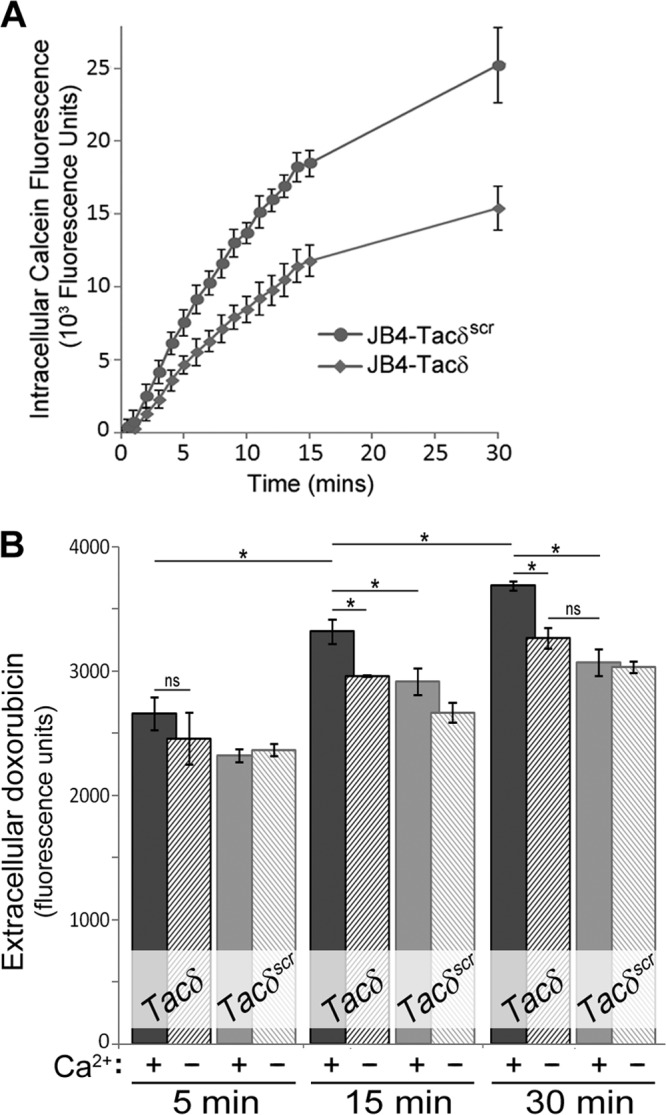
Effects of GFFKR expression on drug efflux. (A) JB4-Tacδ and JB4-Tacδscr cells were incubated with the cell-permeant calcein-AM substrate, and fluorescence measurements were made at the indicated times. Nonfluorescent calcein-AM is hydrolyzed to the highly fluorescent calcein by intracellular esterases; thus, the rate of calcein accumulation is an indirect and inverse measure of the cellular efflux rates of calcein-AM. Data plotted are the mean values ± standard deviations for 4 replicate wells. ns, not significant for t = 0 to 2 min; P < 0.002 for t > 5 min. (B) JB4-Tacδ and JB4-Tacδscr cells were incubated with high doses of doxorubicin, washed free, and resuspended in PBS with (+) or without (−) 1 mM Ca2+. At the indicated times, cell-free supernatants were assessed for fluorescence as an indication of doxorubicin efflux from cells. Sampling for the earliest time point was estimated at 5 min. Data plotted are the means ± standard deviations for 3 replicate cell aliquots. *, P < 0.009; ns, not significant.