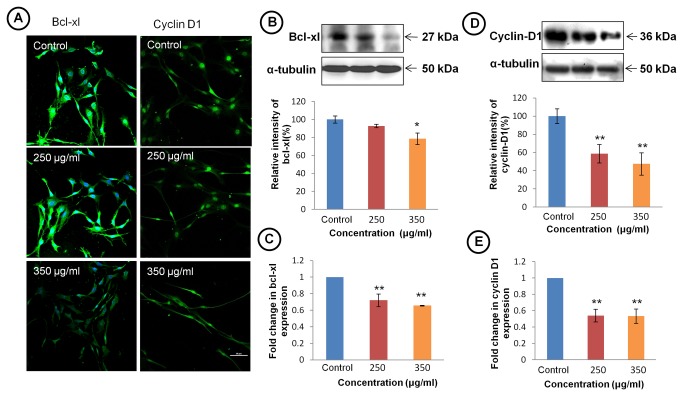
Figure 4. TCE inhibits anti-apoptosis and cell cycle promoting genes.
(A) Confocal images of immunostaining of bcl-xl (left panel) and cell cycle regulator protein cyclin D1 (right panel) in TCE treated and untreated C6 cells (Scale bar- 50 μm). (B) Representative western blot hybridization signals of bcl-xl (upper panel). Histogram (lower panel) representing the relative change in expression of bcl-xl. (C) Histograms representing expression of mRNA of bcl-xl in control and treated cells. Gene expression is represented by ΔΔCt value of bcl-xl after normalising with 18S RNA as endogenous control. (D) Representative western blot hybridization signals of cyclin D1 in TCE treated and control group (upper panel). Histogram (lower panel) represents relative change in expression of cyclin D1. (E) Histograms representing expression of mRNA of cyclin D1 in control and treated cells. Gene expression is represented by ΔΔCt value of cyclin D1 after normalising with 18S RNA as endogenous control. Values are presented as mean ± SEM of at least three independent experiments. ‘*’ (P<0.05) and ‘**’ (p< 0.01) represent statistical significant difference between control and TCE treated groups.

