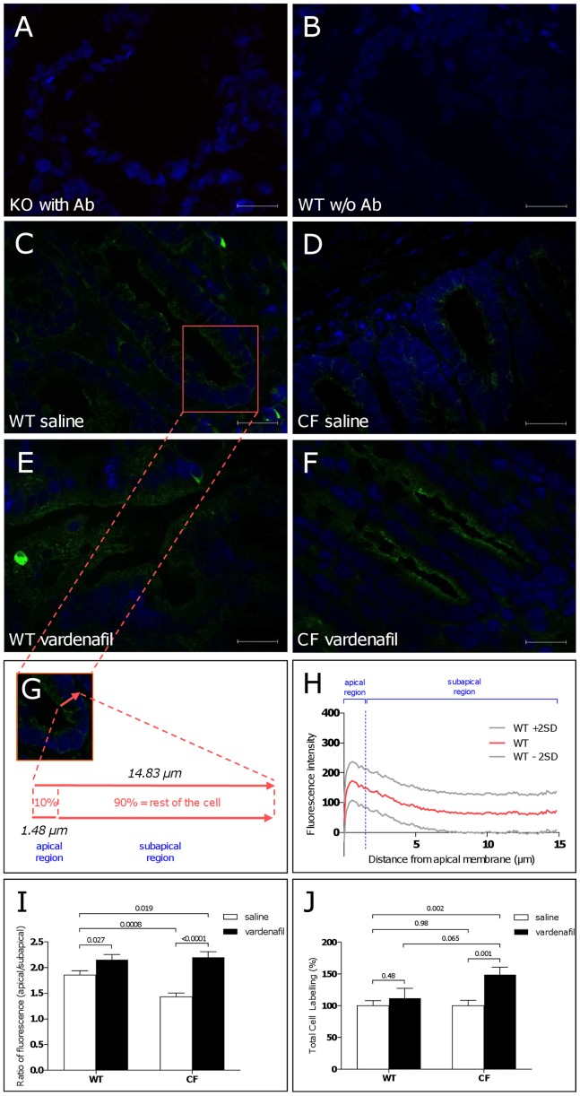
Figure 5. Immunohistochemical localization studies showing absence of labelling of endogenously expressed CFTR in distal colon tissue from a Cftr knockout mouse (A) and a wild-type mouse (B) in the absence of primary anti-CFTR antibody (w/o Ab).
Immunolabelling performed 1 hour after a single intraperitoneal injection of saline (C,D) or 0.14 mg/kg vardenafil (E,F) in crypt colonocytes from a wild-type mouse (C,E) or a F508del-CF homozygous mouse (D,F). Vardenafil treatment (E,F) increased CFTR (green) labelling at the apical membrane compartment. Nuclei (blue labelling) stained by DAPI. Morphometric analysis of crypt cells with measure of the apical and subapical (corresponding to the rest of the cell height) compartments (G). Mean values and upper/lower 95% confidence intervals (±2SD) of scans of the intensity of the CFTR fluorescence signal along a line drawn through the apical to the basal cell borders obtained from 136 crypt colonocytes from saline-treated wild-type mice; the vertical line marks the apical compartment corresponding to the upper 10% of the height of the cell; total area under the curve = 1285 µm.intensity unit; area under the curve of the apical region = 219.6 µm.intensity unit; peak intensity = 172.8 units; distance from apical membrane to peak intensity = 0.555 µm (H). Normalized ratio of apical/subapical fluorescence CFTR signal (I) and total cell labelling (J) in saline-treated and vardenafil-treated mice.

