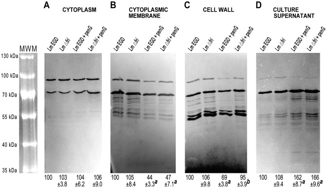
Figure 3. Content of murein hydrolases in various cellular compartments of L. monocytogenes strains.
Zymography analysis of proteins from the cytoplasm (A), cytoplasmic membrane (B), cell wall fraction (C) and culture supernatant (D) were performed for the wild-type EGD strain grown without (Lm EGD) and with (Lm EGD + penG) penicillin G, and the Δ fri mutant strain grown without (Lm Δfri) and with (Lm Δfri + penG) the same antibiotic. Equivalent quantities of proteins isolated from each fraction were subjected to zymographic analysis in SDS-polyacrylamide gels. MWM – prestained Protein Molecular Weight Marker. The relative amounts of murein hydrolases estimated on the basis of the densitometric analysis are shown in the lower panel. Values in each analyzed compartment were normalized to the total intensity of murein hydrolases from the wild-type EGD strain grown without the antibiotic, which was assigned the value of 100%. The presented results are mean values from the analysis of three independent protein isolations ± the standard deviation. a Significant differences following growth in the presence and absence of penicillin G (Student's t-test; P<0.05). b Significant differences between the studied strains (Student's t-test; P<0.05).

