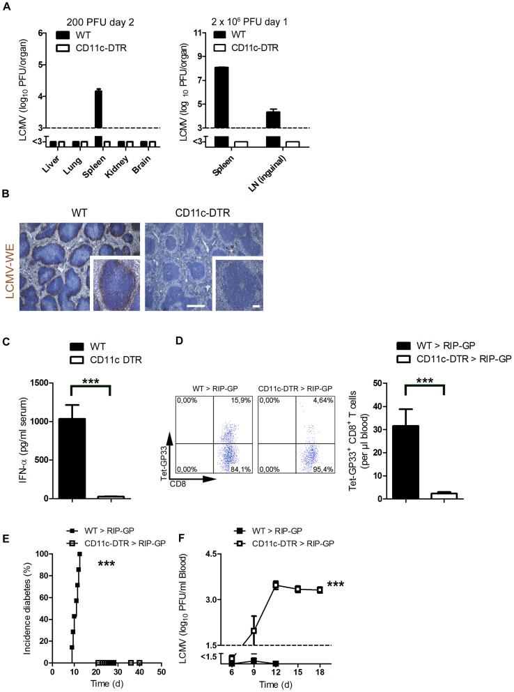
Figure 1. Depletion of dendritic cells blunted early viral replication and prevented autoimmune diabetes.
(A) CD11c-DTR mice and C57BL/6 mice were treated intraperitoneally with diphtheria toxin (30 µg/kg) on day −3. Mice were infected with LCMV (200 PFU) or LCMV (2×10⁶ PFU) on day 0. Viral titers were analyzed at the indicated time points in different organs (200 PFU n = 6 and 2×10⁶ PFU, n = 3). (B) C57BL/6 and CD11c-DTR mice were treated intraperitoneally with diphtheria toxin (30 µg/kg) on day −3 and then infected with LCMV (2×10⁶ PFU) on day 0. After one day, immunohistologic staining for LCMV-NP was performed on spleen sections (n = 3, scale bar main images 500 µm, inlets 100 µm). (C) CD11c-DTR mice and control WT mice were treated with 30 µg/kg diphtheria toxin on day −3. On day 0 mice were infected with 2×10⁶ PFU LCMV. After two days IFN-α was measured in the serum by ELISA (n = 6). (D–F) RIP-GP mice were lethally irradiated and one day later were reconstituted with 107 bone marrow cells from either CD11c-DTR mice or C57BL/6 mice as control animals. Thirty days later, mice were treated intraperitoneally with diphtheria toxin (10 µg/kg) on days −1, 2, 5, and 8 and were infected intravenously with 200 PFU LCMV-WE on day 0. A representative dot plot and the quantification of virus specific GP33+ CD8+ T cells analyzed on day 8 in the blood with FACS analysis is shown (n = 10–14, D). The incidence of diabetes was determined by measuring serum glucose concentrations after LCMV infection (n = 7–11, E). Virus titers were analyzed in the blood at different time points after infection by plaque assay (n = 5–11, F). *** P<0.001 (Student's t-test) (C and D), Log-rank (Mantel-Cox) (E) two-way analysis of variance (ANOVA) (F).

