
Figure 7. Apotome imaging after 7 days of biofilm development on a glass surface of a mixture of A) AG L. pneumophila Lens or B) MG L. pneumophila Lens expressing Ds-Red marker (in red) or GFP marker (in green) and polysaccharides (in blue).
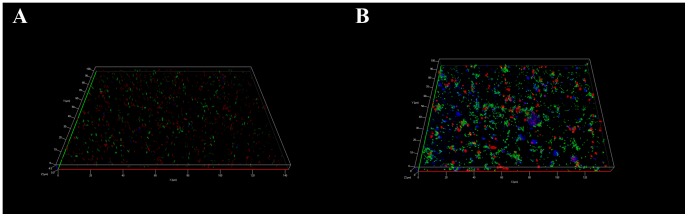
Pictures are representative of 3 independent experiments.