
Figure 10. Apotome imaging after 7 days of biofilm development on a glass surface of a A) L. pneumophila Lens treated with supernatant of AG L. pneumophila Lens or B) with supernatant of AG L. longbeachae.
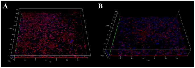
Bacteria are in red and polysaccharides appear in blue. Pictures are representative of 3 independent experiments.