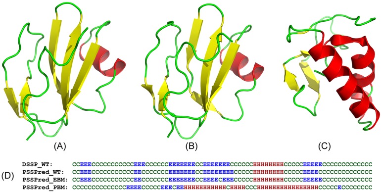
Figure 3. Illustration of protein design on the soluble human CD59.
(A) X-ray structure of the target protein. (B) I-TASSER model on the EBM designed sequence. (C) I-TASSER model of the PBM designed sequence. (D) Secondary structure of the target assigned by DSSP, in comparison to that predicted by PSSpred on the target (PSSPred_WT), the EBM (PSSPred_EBM), and the PBM designed sequences (PSSPred_PBM). ‘E’ stands for sheet, ‘H’ for helix and ‘C’ for coil.

