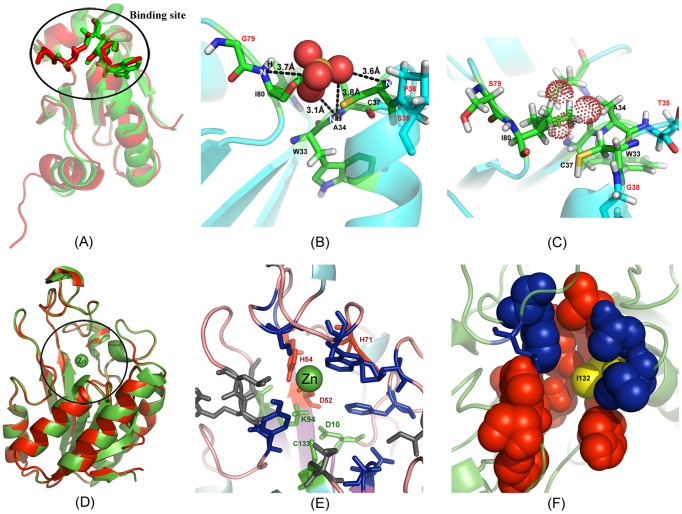
Figure 6. Illustrative examples of the EBM design on M. tuberculosis proteins.
(A) Superposition of I-TASSER model of the EBM sequence (green) on the target structure from the thioredoxin C (red) with a RMSD 2.52 Å. (B) Sulfate ion binding with the designed protein where ion-protein hydrogen bonds are highlighted by dashed lines. (C) The EBM binding site analogous position on the target indicates that it cannot accommodate the sulfate ion (dotted sphere) due to steric overlaps. (D) Superposition of the PZAase protein (red) and the I-TASSER model on the EBM sequence (green) with a RMSD 0.28 Å. (E) Active site residues of PZAase as represented in sticks. Triad (green) and Zn2+ binding sites (red) are retained in the designed protein. Gray color indicates mutations at the active site. (F) Binding pockets identified by COFACTOR with red spacefill indicating an isochorismic acid binding site and blue the sulfate ion binding site. Y132I mutation in EBM design is designated by yellow. The figure was generated using Pymol and Adobe Photoshop software.

