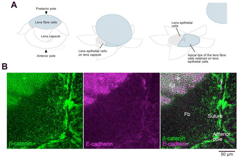
Figure 3.
Preparation of lens whole mounts. A. Diagram showing how the lens capsule is peeled away from the lens fiber cells. Lens epithelial cells remain firmly attached to the lens capsule; however, when the fibers are removed, in some regions, remnants of their apical ends are retained on the apical surface of the epithelial cells. B. β-catenin (green) is present at the margins of both epithelial and fiber cells but E-cadherin (purple) is specific for epithelial cells. In this whole mount, a large region near the anterior pole of the lens retains remnants of the apical ends of lens fiber cells. Abbreviations: Ep, epithelial cells; Fb, fiber cells. Adapted from Sugiyama et al, 2010.

