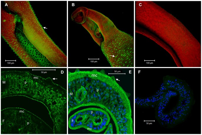
Figure 5. Fluorescence confocal microscopy images showing immunolocalization of SmCD59.1 and SmCD59.2 in whole mount and in transverse sections of S. mansoni adult worms.
(A and B) Confocal projections of SmCD59.1 and SmCD59.2 protein in the tegument of whole mount adult male worms. (D and E) Fluorescence detection of SmCD59.1 and SmCD59.2 in transverse sections of S. mansoni adult worms. (C and F) Negative control, serum from naïve rat. Secondary antibody coupled to Alexa 488 (green) was used for SmCD59 localization. DAPI (blue) was used for nucleus localization (E and F) and Rhodamine Phalloidin (red) was used for actin localization (A, B and C). Arrows – tegument tubercules; M – male; F – female; p – parenchyma; mc – muscle cells.

