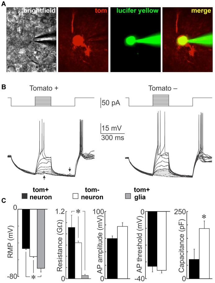
Figure 6. Electrophysiological properties of NG2-glia derived neurons.
A neuron targeted by a patch pipette in a living hypothalamic slice is visible by brightfield, expresses Tom, and is filled with Lucifer yellow during the course of recording (A). Whole cell voltage recordings obtained from a Tom positive (left) and a Tom negative (right) neuron (B). The traces show superimposed voltage responses (lower) to current pulses (upper). Note that both types of cells can fire action potentials. Synaptic potentials are visible in the Tom+ neuron (arrows). Bar graphs compare mean (± SEM) resting and active membrane parameters recorded from excitable Tom+ neurons, conventional neurons (Tom negative neurons) and putative glia (unexcitable Tom+ cells)(n = 4) (C). RMP = resting membrane potential, AP = action potential, *p<0.05.

