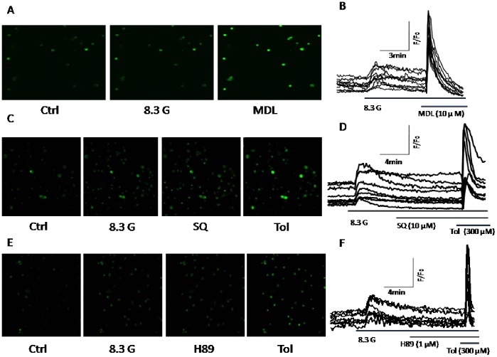
Figure 7. MDL-12,330A increases [Ca2+]i level.
[Ca2+]i was measured from beta cells loaded with Fluo 4-AM using a laser scanning confocal microscope at room temperature. The ratio of fluorescence change F/F0 was plotted to represent the change in [Ca2+]i levels. A, B. cells were treated with 10 µM MDL-12,330A (MDL, n = 10) in the presence of 8.3 mM glucose (8.3G). C, D. Cells were treated with 10 µM SQ 22536 (SQ, n = 9) in the presence of 8.3 G. E,F. cells were treated with 1 µM H89 (n = 8) in the presence of 8.3 G. As a positive control, Tolbutamide (Tol, 300 µM) was applied after SQ 22536 or H89 treatments.

