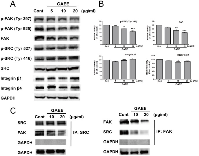
Figure 4. GAEE suppressed FAK signaling in MDA-MB-231 cells.
(A) Immunoblots of FAK signaling proteins and other related proteins after 24 h of treatment with GAEE in MDA-MB-231 cells. Decreased expression of FAK, p-FAK (Y397), and p-FAK (Y925) were shown, while there is no alter on the expression of SRC, p-SRC, integrin β1, and integrin β4. (B) The relative densities of FAK, p-FAK (Y397), integrin β1 and integrin β4 were normalized against GAPDH by densitometric analysis. The values represented as the mean ± SEM of four independent experiments compared with control group. *P<0.05, **P<0.01, ***P<0.001 (one-way ANOVA with Tukey's multiple comparison test) (C) IP-Western confirmation for the disruption of interaction between FAK and SRC after treatment with GAEE.

