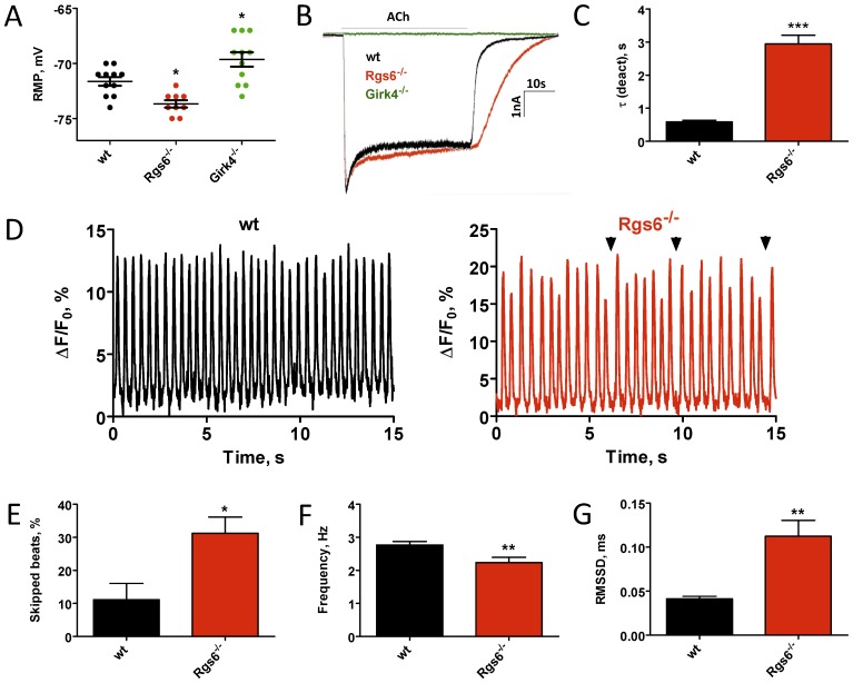
Figure 3. Ablation of Rgs6 reduces excitability of sinoatrial cells and disrupts their automaticity.
A, Resting membrane potential measured immediately after obtaining whole-cell access in wild-type (wt), Rgs6−/−, and Girk4−/− SAN cells. B, Inward currents evoked by application of acetylcholine (ACh, 100 µM) in SAN cells from wild-type (black), Rgs6−/− (red) and Girk4−/− (green, no current) mice. C, Summary of steady-state ACh-induced deactivation kinetics of IKACh in wild-type and Rgs6−/− SAN cells (n = 11–15 cells/genotype). D, Representative traces of spontaneous calcium oscillations recorded from wild-type (black; n = 14) and Rgs6−/− (red, n = 20) SAN cells. Arrows show skipped beats. E, Quantification of SAN arrhythmic events defined as more than 15% change in duration of peak-to-peak interval of spontaneous calcium oscillations in wild-type (n = 11) and Rgs6−/− (n = 17) SAN cells. F, Reduced frequency of spontaneous calcium oscillations recorded in Rgs6−/− SAN cardiomyocytes as compared to wild-type (n = 14–20 cells/genotype). G, Increased variability of spontaneous calcium oscillations in Rgs6−/− SAN cells as determined by increase in RMSSD values (n = 14–20 cells per genotype). Symbols: *P<0.05; **P<0.01; ***P<0.001.

