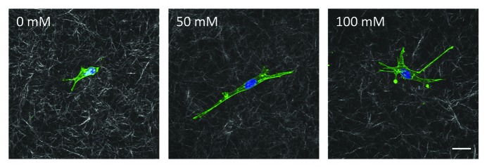
Figure 2. Bovine aortic endothelial cells embedded within glycated collagen gels. Collagen solutions that had been glycated with 0, 50 or 100 mM ribose were neutralized, mixed with endothelial cells and allowed to polymerize. Cells were allowed to spread for 24 h and then were fixed and stained for actin (green) and DAPI (blue). Cells and the surrounding collagen were imaged using confocal microscopy. Scale is 20 μm.
