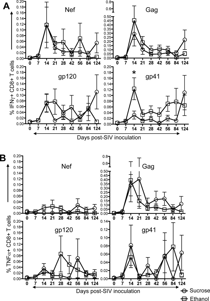
Figure 3.
SIV specific T-cell responses in either ethanol or sucrose treated SIV infected macaques. Intracellular IFNγ responses (A) and TNFα responses (B) were measured against SIV-Nef, SIV-Gag, SIV-gp120, and SIV-gp41 antigens at 9 different time points post SIV infection. Data presented as means ± standard errors of either CD8+IFNγ+ T-cells or CD8+TNFα+ T-cells at different time points of infection. In general, increased IFNγ/TNFα responses were detected in ethanol treated macaques compared to sucrose treated controls in peripheral blood CD8+ T-cells. Asterick (*) indicates statically significant difference between sucrose and ethanol treated SIV infected macaques for the specified time points. Criteria for a positive cytokine response was a two-fold increase in frequency for that specific antigen and cytokine above the medium control culture. All values were subtracted from medium control before analysis.

