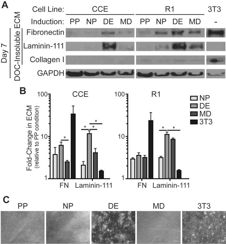
Figure 4. Mouse ESC-derived ECM is distinct from 3T3 fibroblast-derived ECM and is affected by induction medium.
Mouse ESCs (CCE and R1) and 3T3 fibroblasts were cultured for 7 days in pluripotency (PP), neural progenitor (NP), definitive endoderm (DE), mesoderm (MD), or 3T3 fibroblast medium. (A) A DOC-solubility assay was used to isolate the DOC-soluble (cell lysis) fraction from the DOC-insoluble extracellular matrix. Both fractions were separated by SDS-PAGE and blotted for GAPDH and extracellular matrix proteins, respectively (top). (B) The fold-change in expression of the indicated extracellular matrix protein was quantified relative to the pluripotency (PP) condition and the GAPDH content in the DOC-soluble fraction (right) (mean ± standard error from 3 independent samples). (C) The cell-derived extracellular matrices from each culture condition were decellularized and imaged by phase-contrast microscopy. Scale bar is 50 μm. * p < 0.05 as determined by ANOVA.

