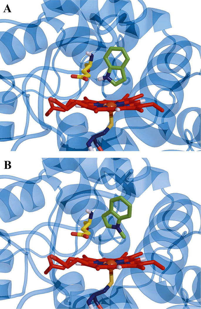
Figure 4.
Ribbon and stick representations of (A) indole and (B) 1-methylindole docking in the active site of CPO. There was only one orientation of indole and 1-methylindole found in docking. The indole and 1-methylindole are colored green. Heme is shown as red sticks, and Glu 183 is shown as yellow sticks.

