
Figure 1.
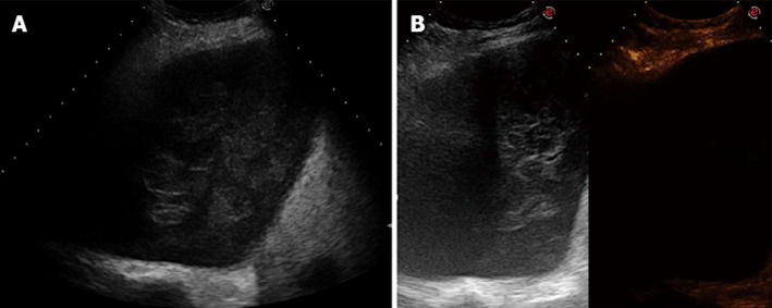
Absence of arterial enhancement. A: transverse sonogram of the right chest wall in the sixth posterior intercostal space showing an inhomogeneous mass in the pleural space; B: transverse contrast-enhanced sonogram of the mass showing complete absence of enhancement in the arterial phase. Final diagnosis was haemothorax with a large clot.