
Figure 3.
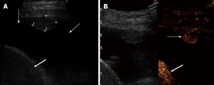
Delayed arterial enhancement. A: Tranverse sonogram of the left chest wall in the seventh intercostal space along the mid-axillary line showing an isoechoic pleural nodule, pleural effusion (thin arrows), and spleen (large arrow); B: Transverse contrast-enhanced sonogram in the arterial phase showing inhomogeneous enhancement of the nodule (thin arrow) that occurs contemporaneously to the enhancement of the spleen (large arrow) (left side of the split-screen); the enhancement of the nodule is less marked than that of the spleen. Final diagnosis was pleural metastasis from colon cancer.