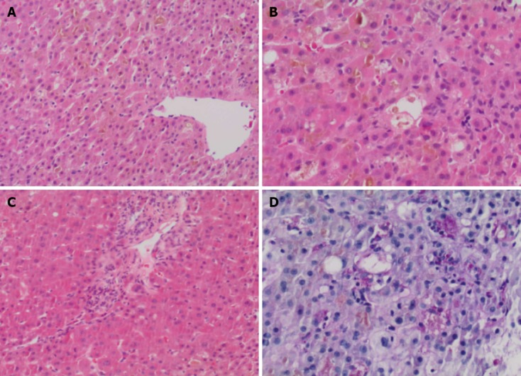
Figure 1.

Histopathological changes. A: The liver biopsy showed marked cannalicular cholestasis, predominantly in a pericentral (zone 2 and 3) distribution; B: Numerous large intracannicular bile thrombi were present throughout the hepatic parenchyma, associated with early foamy degeneration, hepatocyte rosettes and patchy cytoplasmic condensation and eosinophilia. Lobular inflammation, as is the norm in cholestatic hepatitis, were minimal within the lobule; C: The portal tracts showed only minimal inflammation, without any evidence of bile duct injury or loss and there was no interface hepatitis present; D: Large numbers of ceroid laden macrophages were identified on PASD stain in keeping with increased hepatocyte turnover. Magnification: (A, C, D) x 40; (B) x 200.
