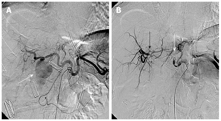
Figure 2.

A 56-year-old man presented with massive bleeding from surgical drains after choledocholithotomy due to common bile duct stone. A: Selective celiac arteriography showed a pseudoaneurysm (arrow) arising from the right hepatic artery; B: Selective celiac arteriography after embolization with microcoils and gelfoam demonstrated disappearance of the pseudoaneurysm. Embolic agents were inserted proximally (white arrow) and distally (black arrow) to the origin of the pseudoaneurysm.
