Figure 6.
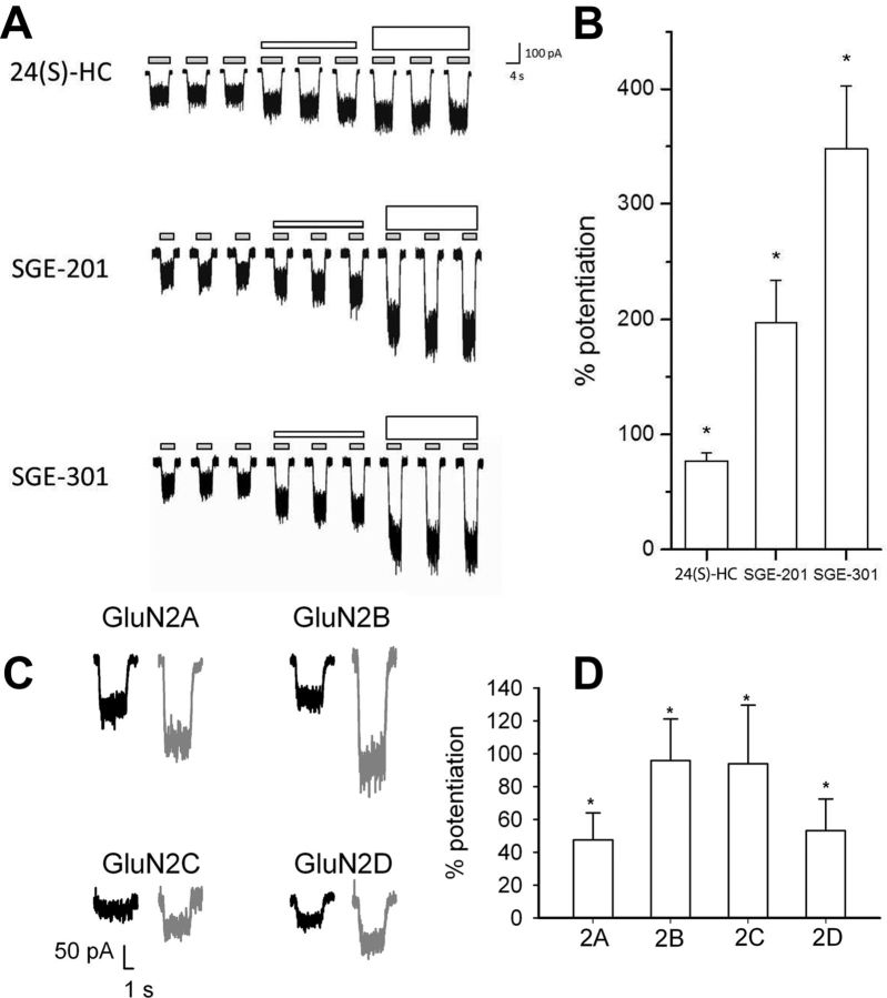
Potentiation of recombinant receptors suggests little or no subunit selectivity. A, HEK-293 cells stably expressing human GluN1–3 and transiently expressing human GluN2A were activated with NMDA (30 μm) and glycine (5.0 μm) (small gray bars). After determining the baseline response to NMDA and glycine, a test compound (as indicated) was added at 0.1 μm (short white bars) or 1 μm (tall white bars). B, The mean (± SEM) percentage potentiation (by 1 μm test compound) above NMDA and glycine alone is plotted. Asterisks denote a significant difference from baseline (p < 0.05). C, Sample traces from HEK cells transiently transfected with GluN1a plus each of the indicated GluN2 subunits. Potentiation of 10 μm NMDA currents (0.5 μm glycine) by 0.2 μm SGE-201 is shown (gray traces of each pair). D, Each subunit combination exhibited significant potentiation by SGE-201 above baseline (asterisks), but no significant difference in potentiation values among subunits was detected.

