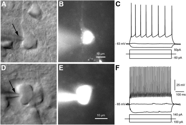
Figure 2.
Identification of Pyr cells and PV neurons in slices of visual cortex. Differential interference contrast image of Pyr cell (A) and PV neuron (D) with recording pipette (arrow) filled with Alexa 594 and biocytin (B, E). Current-clamp recording from retrobead-labeled FFV1→LM-projecting Pyr cell (B) shows that a depolarizing current step evokes an adapting (regular spiking) train of action potentials (C). Recording from a tdTomato-expressing PV neuron shows a nonadapting (fast spiking) train of spikes (F).

