
Figure 6.
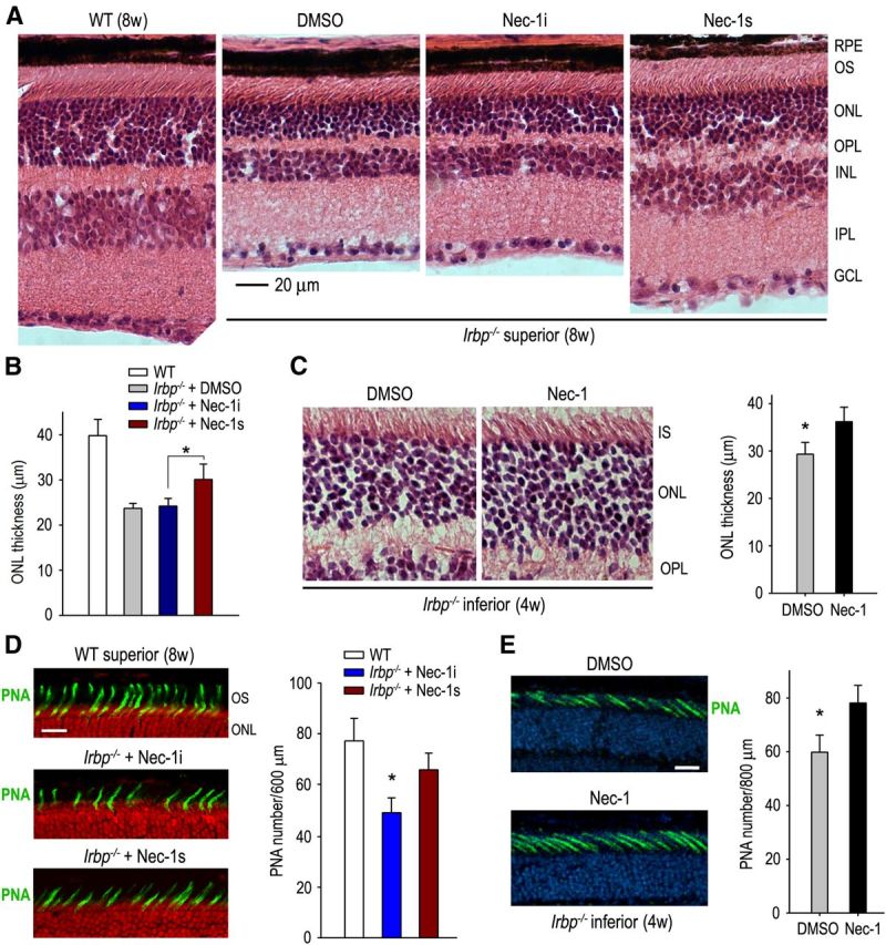
Protection of cone and rod photoreceptors by RIP1 inhibitors in Irbp−/− mice. A, H&E staining of retinal sections from 8-week-old (8w) WT and Irbp−/− mice treated with DMSO, Nec-1i, or Nec-1s. GCL, Ganglion cell layer. B, Histogram showing the average thickness of the ONL in superior retinas from 8w-old WT and Irbp−/− mice treated with the indicated reagents. Asterisks indicate statistically significant differences between mice treated with Nec-1i or Nec-1s (p < 0.05). Error bars denote SD (n = 6). C, H&E staining of retinal sections of 4w-old Irbp−/− mice treated with DMSO or Nec-1. The average thickness of the ONL in the inferior retinas of the mice is shown in the histogram. Asterisks indicate significant differences between the two treatments (p < 0.05). Error bars denote SD (n = 6). IS, Inner segment of photoreceptor. D, PNA staining (green) of the superior retinas of 8w-old WT and Irbp−/− mice treated with Nec-1i or Nec-1s. Nuclei were counterstained with propidium iodide (red). Numbers of PNA-positive cone matrix sheathes in a 600 μm width region of the superior retinas are shown in the histogram. Scale bar, 20 μm. E, PNA staining of the inferior retinas of 4w-old Irbp−/− mice treated with DMSO or Nec-1. Nuclei were counterstained with DAPI (blue). Numbers of PNA-positive cone sheathes in 800 μm width region of the mice inferior retinas are shown in the histogram. Scale bar, 10 μm.