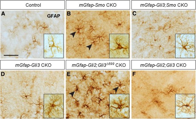
Figure 9.
Unattenuated GLI3R levels alter astrocyte function. A–C, IHC staining of sections of adult cortex for GFAP shows that mGfap-Smo CKO astrocytes exhibit a partial reactive astrogliosis phenotype (A, B), whereas removal of Gli3 in mGfap-Smo CKOs rescues astrocyte morphology and drastically reduces the population of GFAP-overexpressing glia in the cortex (C). D–F, No obvious change in the morphology of GFAP-expressing astrocytes is observed in mGfap-Gli3 CKOs (D) or in mGfap-Gli2;Gli3 double CKOs (F), whereas expression of GLI3R in the absence of GLIA (mGfap-Gli2;Gli3Δ699 CKOs) recapitulates the astrocyte hypertrophy and GFAP overexpression observed in mGfap-Smo CKOs (E). A–F, Insets, Example of cortical astrocyte morphology in the various mutants and control. Scale bar: (in A) A–F, 50 μm.

