Figure 1.

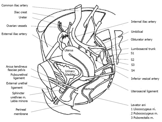
Sagittal section of female pelvis illustrating the position of the sphincter urethrae muscle in relation to adjacent structures and associated neurovascular components. Adapted from reference [21].

Sagittal section of female pelvis illustrating the position of the sphincter urethrae muscle in relation to adjacent structures and associated neurovascular components. Adapted from reference [21].