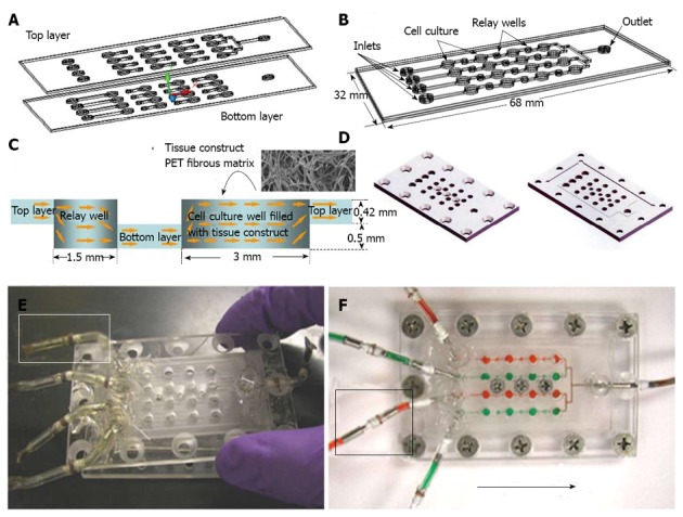
Figure 4.

Perfusion bioreactor array for cell-based assay. A: Schematic drawing of the device composed of top and bottom layers; B: Perspective view of the assembled device to form microscale channels and wells; C: Design of individual cell culture well and relay well formed by two layers, with cell culture well that can be filled with any modular tissue engineering scaffold such as PET fibrous matrix; D: Perspective drawing of the top and bottom frames for frame-assisted assembly; E: Photograph of device in assembly with each inlet connecting to a flexible connector capped to prevent contamination (highlighter window); F: Photograph of assembled device at work with each inlet connected to an external tubing through a flexible connector (highlighter window). Reproduced from reference Wen et al[14].
