
Figure 3.
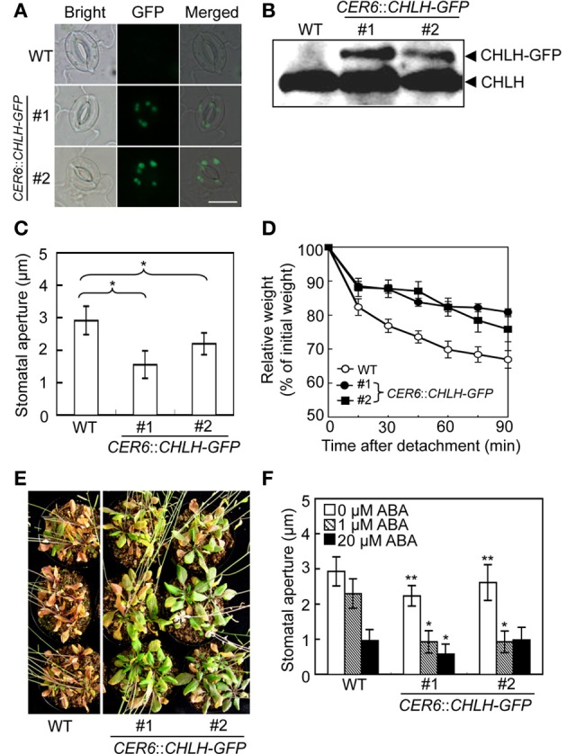
Phenotypic analysis of CHLH transgenic plants. The phenotypes of two independent CER6::CHLH-GFP plants were compared with that of WT plants. (A) Typical bright-field and GFP fluorescent images of stomata in the abaxial epidermis of rosette leaves. Scale bar = 10 μm. (B) Immunoblots using anti-CHLH antibody showing CHLH-GFP and endogenous CHLH in GCPs. (C) The stomatal aperture in 4-week-old plants exposed to light conditions at zeitgeber time (ZT) 4. Epidermal fragments were isolated from rosette leaves and apertures measured immediately. Data represent the means of 25 measurements ±SDs. Pairs for Student's t-test are indicated with brackets (*P < 0.01). (D) Kinetics of fresh weight changes in detached rosette leaves from 4-week-old WT plants (open circles) and CER6::CHLH-GFP #1 (closed circles) and #2 (closed squares). The relative weights of leaves are presented (±SD) as percentages of initial weights, being the weights of rosette leaves immediately after detachment from plants (n = 6). (E) Drought tolerance in WT and CHLH-GFP transgenic plants. Plants grown in soil for 3 weeks were subjected to drought stress by withholding water for 18 days. (F) Effect of ABA on the stomatal aperture of WT and CHLH-GFP transgenic plants. Epidermal fragments isolated from rosette leaves of 4-week-old plants exposed to light at ZT 4 were treated with ABA at the indicated concentrations for 2.5 h under BL superimposed on background red light. The stomatal apertures in the abaxial epidermis were measured microscopically. Data represent the means of 25 measurements ±SDs. Asterisks indicate significant differences between wild type and each CHLH-transgenic plants under 0 μM ABA (*P < 0.05; Student's t-test). The experiment was repeated three times on different occasions with similar results.