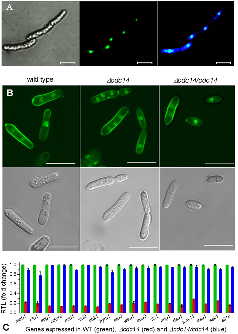
Figure 1. Disruption of cdc14 caused cytokinesis defect in B. bassiana.
(A) Microscopic images of transgenic hyphal cells expressing the fused protein eGFP::Cdc14 in SDB at 25°C. Left: bright image of the cells stained with DAPI. Middle: fluorescent image of the expressed eGFP in nuclei at the excitation/emission wavelengths of 488/507 nm. Right: fluorescent image of the DAPI-stained Cdc14 in nuclei at the excitation/emission wavelengths of 358/360 nm. (B) Bright (upper) and fluorescent (lower) images of hyphal cells stained with both DAPI and calcofluor white (a stain specific to cell wall). Note that cytokinesis was normal (with one or two nuclei per cell) in wild type but abnormal (with three or more nuclei per cell) in Δcdc14. Scale bars: 10 μm. (C) Relative transcript levels (RTL) of 18 cytokinesis-associated genes in Δcdc14 and Δcdc14/cdc14 versus wild type grown for 3 days in SDB at 25°C. Error bars: SD from three cDNA samples assessed via qRT-PCR with paired primers (Table S1).

