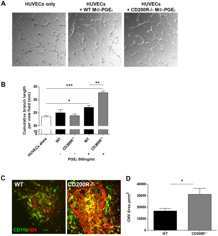
Figure 3. Enhanced pro-angiogenic phenotype of CD200R-deficient macrophages promotes angiogenesis.
(A) Representative images of HUVEC cells cultured alone, or in the presence of WT or CD200R−/− macrophages stimulated with PGE2 for 24 h in advance. (B) Cumulative branch length per field of HUVEC cells cultured alone, or in the presence of WT or CD200R−/− macrophages stimulated, or not, with PGE2. Data are presented as mean ± SEM, n = 3, *p < 0.05; **p < 0.005; ***p < 0.0005 between groups. (C) Representative confocal images of RPE/choroid flat-mounts from WT and CD200R−/− mice at day 7 post-laser application, were immunostained with CD11b (green) and Isolectin B4 (IB4) (red). (D) Mean CNV area calculated from confocal images. Data presented as mean ± SEM, n = 26 lesions for each strain; *p < 0.05.

