
FIG 1 .
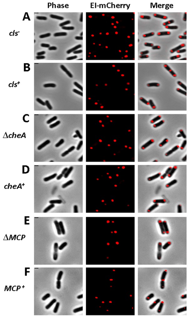
Polar localization of EI does not depend on cardiolipin and the chemotaxis complex. Images showing EI-mCherry distribution in WC3899 E. coli cells mutated in the cardiolipin synthase (cls) gene (A) compared to the parental W3899 cls+ strain (B) and in AW546 ∆cheA E. coli cells deleted for the cheA gene (C) or in UU2612 E. coli cells deleted of all chemotaxis receptors (E) compared to their respective parental strains AW546 and RP437 (D and F, respectively). The mCherry fusion protein was observed by fluorescence microscopy (red), and the cells were observed with phase microscopy (gray). Also, overlays of the signals from the fluorescence and phase microscopy are shown (merge). Scale bar corresponds to 1 µm.