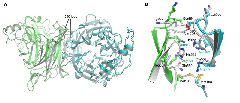
FIG 3 .
Introduction of a Q559R mutation weakens the HN-HN dimer interaction. (A) Structural comparison between HPIV3-HNH552 (reference strain) and HNQ552/R559. The HPIV3 H552 (reference strain) is shown in gray, and the two HNQ552/R559 protomers that form a noncrystallographic dimer are superimposed on the reference strain and are in green and cyan, respectively. The mutations induce local conformational changes on the dimer interface. Most notably, HNQ552/R559 displays a slightly wider separation across the dimer interface near the 550 loop region. (B) Dimer interface around the 550 loop in HNH552 and HNQ552/R559. The two HNQ552/R559 protomers in the noncrystallographic dimer are in green and cyan, respectively. H552 (from the reference strain) is shown in gray for comparison.

