
FIG 1 .
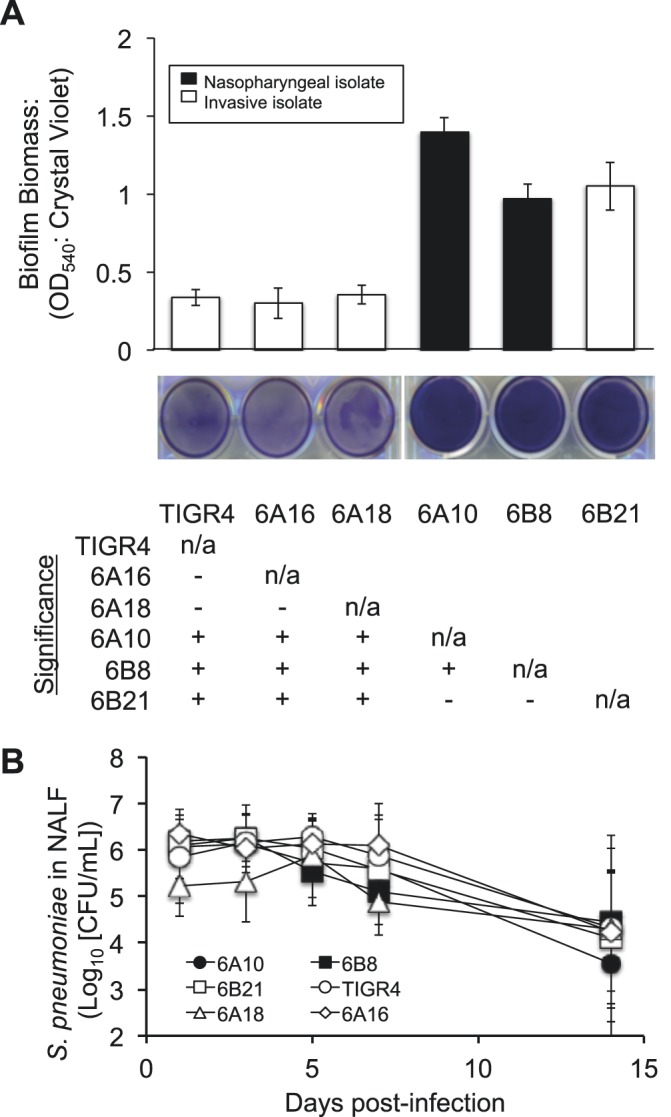
In vitro biofilm formation by S. pneumoniae clinical isolates and corresponding rates of colonization. (A) Mean biomass of biofilms formed by clinical isolates on 6-well polystyrene plates after 18 h of growth, as measured by crystal violet staining (n = 4/strain). Accompanying representative images of stained wells are provided beneath the graph. The table represents statistical significance between strains (+, significance; −, no significance) grown on untreated plates as determined by one-way ANOVA. n/a, not applicable; OD540, optical density at 540 nm. (B) CFU determination of S. pneumoniae in NALF collected from colonized mice at days 1, 3, 5, 7, and 14 postinfection. No significant differences were observed between strains on any given day as tested by one-way ANOVA (n = 9 to 12/cohort).