Figure 1B .
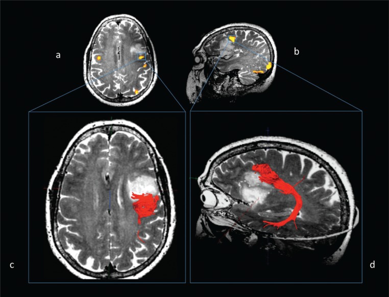
Same case. Functional connectivity: fMRI (a, b) and MR diffusion tractography (c, d).
Left arcuate fasciculus reconstruction using fMRI clusters of activation in the dorsolateral prefrontal cortex as seeding points, evoked during a word generation task and overlaid on T2-weighted images (a, axial view; b, sagittal view). Fibers of the left arcuate fasciculus (in red) overlaid on axial (c) and sagittal (d) T2-weighted images, although strictly adjacent to the posterior margin of the lesion, are dislocated but not infiltrated by the tumor.

