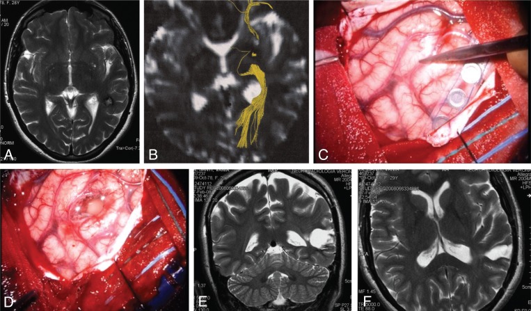
Figure 3 .
Subcortical left temporal cavernous angioma.
The critical surgical issues are: corticectomy in a safe area, safe removal of perilesional gliosis, and sparing of the optic radiations. (A) T2-weighted axial MR scan showing the hypointense subcortical lesion. (B) Diffusion imaging highlighting the optic radiations (yellow). (C,D) Intraoperative pictures showing the site of an essential language area in the middle temporal gyrus (C) and the site of corticectomy in the superior temporal gyrus (D). (E,F) Postoperative T2-weighted coronal and axial MR scans showing the poroencephalic cavity, larger than the size of the cavernous angioma because it encompassed the surrounding gliotic area.

