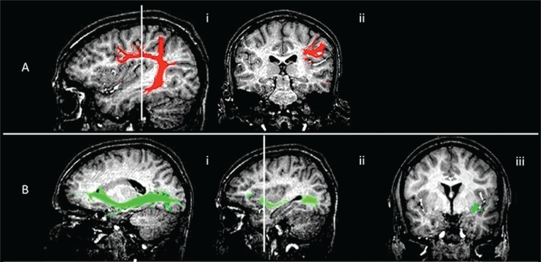
Figure 4 .
Functional imaging showing subcortical surgical landmarks.
MR diffusion tractography anatomy (healthy volunteer). Arcuate fasciculus/superior longitudinal fasciculus and inferior fronto-occipital fasciculus fiber tract reconstruction overlaid on 3D T1-weighted images, showing their relationship with the surgical landmarks.
(A) Arcuate fasciculus. i) sagittal view, anatomy of the tract; ii) coronal view, the tract runs deep in the intersection between the distal sylvian point and the superior sulcus of the insula. This fasciculus runs medially and parallel to the superior longitudinal fasciculus which connects the prefrontal, parietal and temporal cortices.
B) Inferior fronto-occipital fasciculus. i) sagittal view, anatomy of the tract; ii) sagittal reference for the coronal view, at the bottom of the insula, at the level of anterior sulcus, before the ascending part of the tract ; iii) coronal view, at this level the tract is lateral, it then turns medially towards the caudate nucleus.

