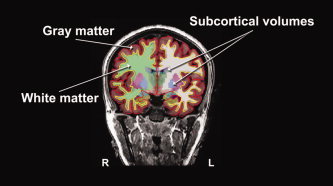
Figure 2.

Calculation of cortical and subcortical volumes with FreeSurfer The pial surface, or gray matter surface (red outline) and the white matter surface (yellow outline) are shown, overlaid over the T1‐weighted image. The segmented cortical volumes are shown in shaded colors. Volumes of the seven subcortical structures of interest were calculated using FreeSurfer's automatic quantification of subcortical structures. Total gray matter volume was calculated by obtaining the volume between the gray and white matter surfaces. Total white matter volume was calculated by subtracting the subcortical and ventricular volumes from the volume bounded by the white matter surface.
