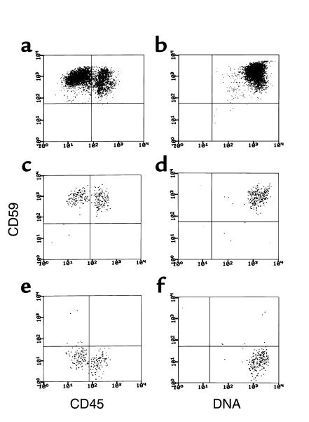
Figure 2.
Flow cytometric analyses of progeny of normal CD34+, patients’ PIGA– CD34+, and patients’ PIGA+ CD34+ cells after 11 days of growth in liquid culture. The cells were stained as in Figure 1. The left (a, c, and e) and right (b, d and f) panels show CD59 vs. CD45 and CD59 vs. DNA, respectively. The upper panels (a and b) show the same patterns for normal cells seen in the preliminary studies. As seen in the middle panels (c and d), the progeny of PIGA+ cells were uniformly CD59+, and as seen in the lower panels (e and f), those of PIGA– cells were uniformly CD59–. For the normal cells, 10,000 gated events were collected based on forward and side scatter and cells were analyzed for CD59 and CD45 or CD59 and DNA. Due to lower available cell numbers, for the PIGA-mutated and -nonmutated patient cell populations, about 1,000 gated events were collected.

