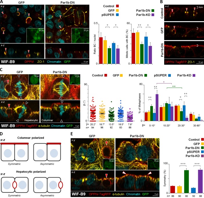
Figure 3.
Par1b depletion in WIF-B9 cells promotes columnar polarity, x-z spindle alignment with the basal domain, and symmetric cell divisions. WIF-B9 cells (A) or DPPIV-TagRFP expressing WIF-B9 cells (B, C, and E) were transduced with adenoviruses expressing either GFP-tagged dominant-negative Par1b (Par1b-DN) or a shRNA-Par1b construct (Par1-KD) or their respective controls (GFP) or (pSUPER-GFP), then fixed and stained for the apical protein (DPPIV), Zonulae occludens 1 tight junctions (ZO-1; A and B), or microtubules (α-tubulin; C and E) and chromatin. GFP expression was monitored (A, C, and E; insets, left panels). The amount of BC-like lumina in interphase or mitotic cells (A; right panels), the β angle (C, right panels), and the distribution of the BC-like lumen or the apical domain into one or both daughters in cytokinetic cells (E, right) were quantified. The shift from larger to smaller β angles caused by Par1b inhibition was statistically significant for β angles in the 0–10° (59.5 ± 8.3%) category compared with the 10–20° (32.3 ± 9.6%) category (Par1b-KD) and between the 10–20° (39.7 ± 2.3) category and 20–30° (21.7 ± 5.6) category for Par1b-DN cells. (D) Modes of cell division encountered in cells with columnar and hepatocytic polarity (depicted in the x-z dimension). Red arrowheads, BC-like lumina; black arrowheads, predicted cleavage furrow plane; white arrowheads, apical domain; yellow arrowheads, chickenwire belt of tight junctions. Error bars indicate ± SEM (dot graphics) or + SD (bar graphics). *, P ≤ 0.05; **, P ≤ 0.01; ***, P ≤ 0.001.

