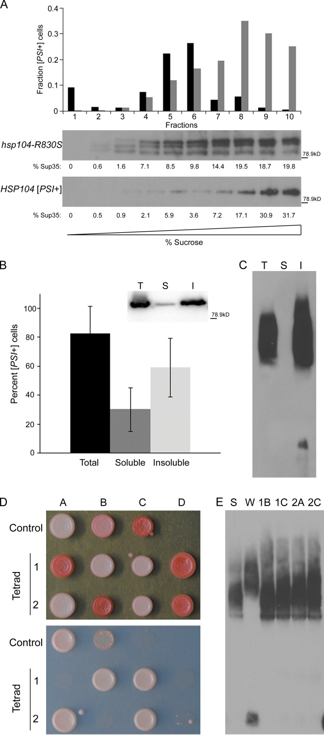
Figure 4.
Soluble oligomeric [PSI+] propagons are infectious and maintain the prion variant structure. (A) Fractions of [PSI+] and hsp104-R830S cryptic [PSI+] lysates separated on a linear sucrose gradient by a 22-h ultracentrifugation step were analyzed by Western blot and the amount of Sup35 (presented as the percent of soluble Sup35) in each fraction was quantified by ImageJ (National Institutes of Health) and is indicated below each fraction in the Western blot. Equal volumes of each fraction were transformed into [psi−] cells. The fraction of infected [PSI+] cells obtained from each gradient fraction is indicated for hsp104-R830S (black) and HSP104 (gray) cells. The fraction of infected [PSI+] cells was generated by compiling the infectivity data from four (HSP104) or five (hsp104-R830S) separate sucrose gradients and transformations. Sup35 in hsp104-R830S lysates appears to be more susceptible to proteolysis and frequently shows degradation products. Protein molecular weight markers (kD) are indicated. (B) Total, soluble, and insoluble fractions of HSP104 [PSI+] lysates from sedimentation analysis were transformed into [psi−] cells. The relative infectivity of the total, soluble, and insoluble fractions was calculated as above and is indicated. (Inset) A Western blot for Sup35 in total (T), soluble (S), and insoluble (I) fractions from one sedimentation assay in this experiment with protein molecular weight marker (kD). Of the thousands of red colonies resulting from transformation of the soluble and insoluble fractions from wild-type [psi−] cells as a negative control, further analysis verified that 48 from each fraction were [psi−] in each of three independent experiments. (C) Total (T), soluble (S), and insoluble (I) fractions of HSP104 [PSI+] lysates from sedimentation analysis were analyzed by SDD-AGE. The soluble fraction is from the top of the supernatant to prevent contamination from the pellet. This experiment was repeated three times. (D) Two representative tetrads from the mating of hsp104-R830S cryptic [PSI+] to [psi−] were spotted on YPD (top) and SD-Ade plates (bottom) to assess the level of nonsense suppression. Strong [PSI+] (Control A), weak [PSI+] (Control B), and [psi−] (Control C) strains were spotted for reference. (E) The lysates of the four white haploids from the tetrads in C were analyzed by SDD-AGE and compared with strong (St) and weak (Wk) [PSI+] controls.

