Figure 3.
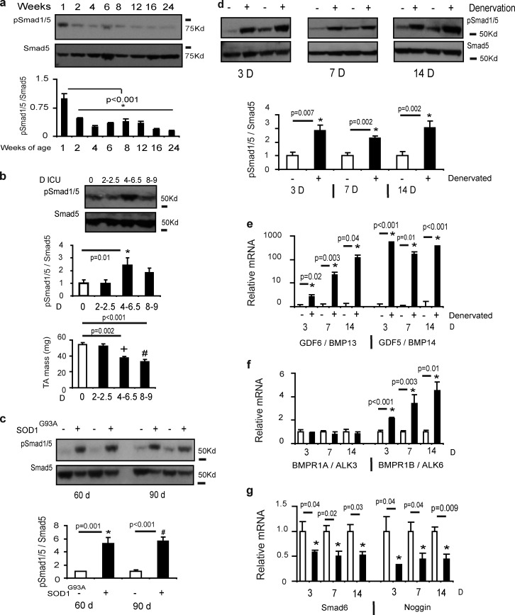
Endogenous BMP signaling is altered in models of skeletal muscle growth and wasting. (a) TA muscles were collected from neonatal and young adult mice and the phosphorylation of Smad1/5 was determined by Western blotting (*, P < 0.001 vs. control). n = 3 per time point. (b) Rats subjected to intensive care immobilization (n = 3–6 per treatment) displayed muscle atrophy with increased phosphorylation of pSmad1/5 (*, P = 0.01 vs. control muscles; +, P = 0.002 vs. control muscles; #, P < 0.001 vs. control muscles). (c) Smad1/5S463/465 phosphorylation was assessed in mice (n = 5 per treatment) with a SOD1G93A mutation that displays symptoms akin to amyotrophic lateral sclerosis patients. At a time point before the onset of muscle atrophy (60 d; *, P = 0.001 vs. control) and at a point of advanced pathology (90 d; #, P < 0.001 vs. control) Smad1/5 phosphorylation was increased. (d) Smad1/5S463/465 phosphorylation was assessed by Western blot 3 d (n = 5 per treatment; *, P = 0.007 vs. control), 7 d (n = 6 per treatment; *, P = 0.002 vs. control), and 14 d (n = 5–6 per treatment; *, P = 0.002 vs. control) after excision of a portion of the peroneal nerve supplying the TA muscle in wild-type mice. (e–g) mRNA expression of GDF6 (BMP13), GDF5 (BMP14), BMPR1A, BMPR1B, Smad6, and Noggin was assessed by RT-PCR (n = 4–6 per treatment; see specific time points for p-values). Gene expression was analyzed using the ΔΔCT method of analysis and expression was normalized to 18S. Data are presented as means ± SEM.

