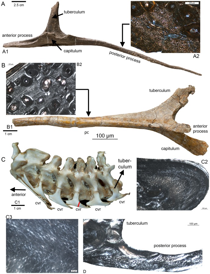
Figure 2. Longitudinal fibers in the posterior processes of cervical ribs of some archosaurs.
A1) Cervical rib from the mid-neck region of a cf. Diplodocus sp. (Sauriermuseum Aathal, Aathal SMA HQ2) in ventromedial view. A2) Histological details of the posterior process of the cervical rib of a cf. Diplodocus sp. (SMA HQ2-D) in polarized light showing dense longitudinally running fibres between scattered secondary osteons. Note the diamond shape of the perpendicular cut longitudinal fibres. The fibres are surrounded by a sheath, which appears here mainly in white (see also Klein et al. 2012). B1) Cervical rib from the sauropodomorph Plateosaurus engelhardti (STIPB R 620) in ventrolateral view. B2) Histological details of the posterior process of the cervical rib of Plateosaurus engelhardti in polarized light showing dense longitudinally running fibers between scattered secondary osteons. C1) Neck from Alligator missisipiensis (STIPB R 599) in lateral view, exhibiting the cervical ribs still attached to the cervicals. In lateral view is only the dorsally located tuberculum visible. cvr = cervical rib. C2) Histological details of the posterior process of a mid-cervical rib of Alligator missisipiensis in polarized light showing dense longitudinally running fibers. C3) Enlargement of the same section, showing longitudinal running fibers. The red line on the posterior process of the mid-cervical rib marks the histological sampling location shown in C2 and C3. D) Histological sample of a posterior process of a mid-cervical rib of an ostrich (Struthio camelus, STIPB R 621) in lateral view and in polarized light showing longitudinally running fibers.

