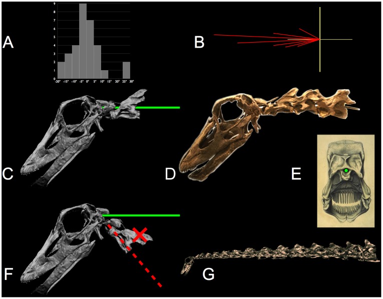
Figure 22. Inner ear orientation is consistent with subhorizontal sauropod necks.
The lateral semicircular canal (LSC) is approximately horizontal in alert birds. The orientation α (see text) is plotted for 32 species of birds [111: fig 7a] as a conventional histogram (A) and polar histogram (B) with 5° intervals (c.f. expanded-scale plot in [132: fig. 2]). When a sauropod cranium is similarly oriented (α=+5°), the rostrum slopes downward (by −15° in Camarasaurus lentus and by −37° in Diplodocus longus) [44], [135]. The LSC also constrains the slope of the neck cranially. The neural canal passing through the atlas-axis is collinear with the foramen magnum, as illustrated by the solid green line in C and the physical armature in the original specimen (D) of Kaatedocus siberi, SMA 0004 [133] – see also the location of the foramen magnum (indicated in green) in the posterior view (E) of Diplodocus [134]. Consequently, with the cranium oriented relative to gravity as indicated by the LSC, and with the cranio-cervical joint undeflected, the anterior neck is roughly horizontal [44]. Taylor et al. [12], however, misinterpreting the anatomy, suggest “… the foramen magnum and occipital condyle are [both] at a right angle relative to the long axis of the skull …” so that the atlas-axis inserts posteroventrally to the cranium, and consequently they falsely conclude the anterior neck ascends steeply as indicated by the red dashed line in F, from [12: fig. 4]; they figured an even steeper neck for Camarasaurus. But properly interpreted, the anatomy of the occiput, the atlas-axis, and the LSC, together with observations of habitual head orientating in the EPB, supports the interpretation that the necks were habitually subhorizontal cranially in diplodocids (E) and camarasaurids (as depicted in Figure 23) [44]. The digital reconstruction (C, F, and G) is based on data courtesy Andreas Christian and Gordon Dzemski. Photo (D) by the author. Supplemental material: Movie S9. A turntable movie depicting the spinal cord (red) entering the foramen magnum of Kaatedocus siberi.

