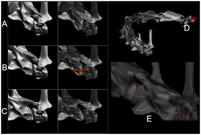
Figure 24. Digital articulation.
CT data of individual vertebrae of a recent giraffe Giraffa Camelopardalis are articulated in Autodesk Maya [108]. Cervical vertebra C7 pivots about a center of rotation that closely corresponds to the center of curvature of the roughly hemispherical condyle of T1, confirmed by exploratory manipulation and adjustment, resulting in close intervertebral separations as reported in [15] (see red arrows). In A–C, by alternating between opaque and transparent one can observe osteological bracing dorsiflexion (A) and the ZSF at the limit of ventriflexion. With all intervertebral joints adjusted (D–E), the articulated neck approximates the range of motion observed in life (see also Figures 11, 12). This method applies equally to the similarly opisthocoelous vertebrae [30]–[32], see Figure 25. CT data provided courtesy American Museum of Natural History.

